The problem
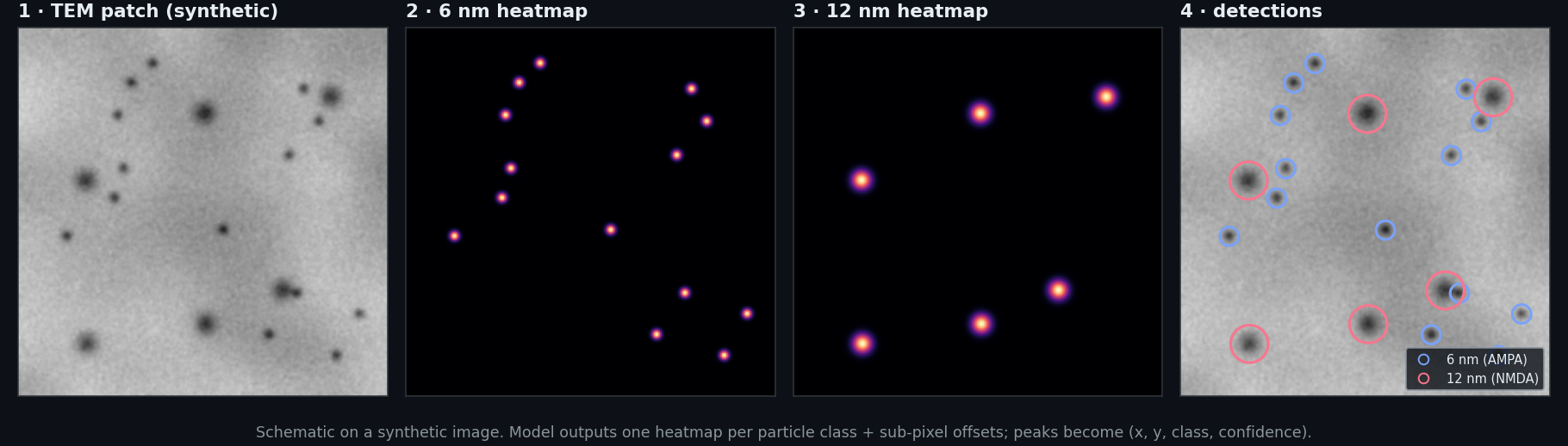
To see where receptors sit at a synapse, neuroscientists tag them with tiny gold beads and image the tissue with a transmission electron microscope. Then someone counts every bead by hand. They're a few pixels wide in a 2048×2048 image, on noisy tissue, and make up well under 0.1% of the pixels.
How it works
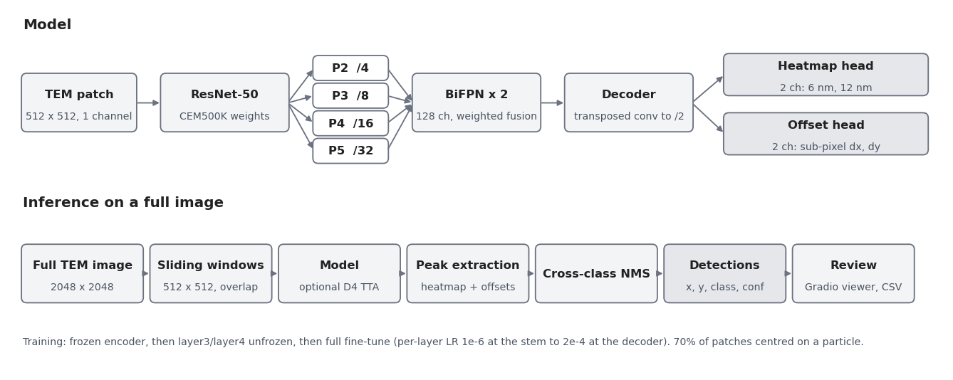
The backbone starts from CEM500K, self-supervised on about 500,000 electron microscopy images, instead of ImageNet photos. A stride-2 output and a BiFPN keep a 6 nm bead big enough for clean peak extraction, and focal loss with particle-centered patches handles the imbalance, where plain BCE collapses to all background.
Limitations
Ten annotated images, 453 hand-labeled particles, each fold testing on one held-out image.
- Those ten images come from one lab; it hasn't been tested on other microscopes.
- The F1 values are from the original evaluation, whose metrics file isn't in the repo. An earlier summary figure shows 0.928 on 429 particles.
- The real images belong to the lab that provided them, so the figures here use a synthetic image.